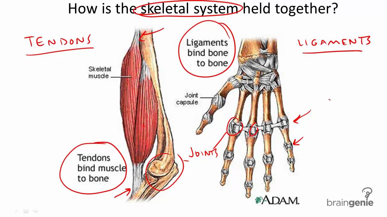
Home » Without Label » Tendon Diagram / Wrist Anatomy Aoa Orthopedic Specialists : The foot diagram has a complex structure made up of bones, ligaments, muscles, and tendons.understanding the structure of the foot is best done by looking at a foot diagram where the anatomy has been labeled.
Tendon Diagram / Wrist Anatomy Aoa Orthopedic Specialists : The foot diagram has a complex structure made up of bones, ligaments, muscles, and tendons.understanding the structure of the foot is best done by looking at a foot diagram where the anatomy has been labeled.
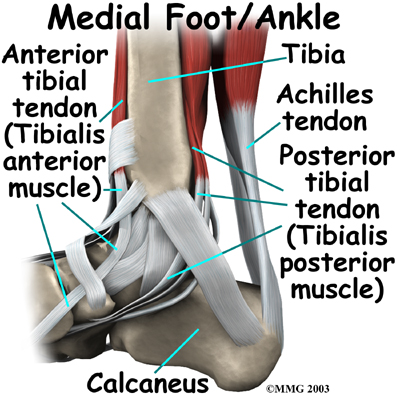
Tendon Diagram / Wrist Anatomy Aoa Orthopedic Specialists : The foot diagram has a complex structure made up of bones, ligaments, muscles, and tendons.understanding the structure of the foot is best done by looking at a foot diagram where the anatomy has been labeled.. Also allows the action of raising up onto toes. The achilles tendon is a tough band of fibrous tissue that connects the calf muscles to the heel bone (calcaneus). The bones of the hip include the femur, the ilium, the ischium, and the pubis. The achilles tendon is also called the calcaneal tendon. This important tendon in the back of the calf and ankle connects the plantaris, gastrocnemius, and soleus muscles to.
When the muscles tighten (contract) arguably, the most important tendon is the achilles tendon, which allows the calf muscles to move. Attaches the calf muscles to the calcaneus, most important muscles for running, jumping, walking etc. Bones, cartilage, ligaments, and tendons. Contusions (bruises) tendonitis (inflammation of a tendon) sciatica (pain from the sciatic nerve) last medically reviewed on june 18, 2015. The anterior cruciate ligament prevents the femur from sliding backward on the tibia (or the tibia sliding forward on the femur).
8 3 4 Tendons And Ligaments Youtube from i.ytimg.com Muscle strains (pulls or tears) muscle cramps; The most commonly accepted description of the mcl is the one originally proposed by milner and soames. Foot anatomy diagram, foot joint diagram, foot sprain diagram, foot tendons and ligaments pain, leg tendon diagram. Allows the action of raising the foot. Hand a hand is a prehensile multi fingered appendage located at the end of the forearm or forelimb of primates such as humans chimpanzees monkeys and lemurs human anatomy for the artist the dorsal hand the dorsal the easiest tendons to identify in the dorsal hand are those of the extensor digitorum muscle its name means extensor of the digits which is Tendons are remarkably strong, having one of the highest tensile strengths found among soft tissues. The hip itself is a ball and socket joint, much like the shoulder.the structures necessary to create this joint are the socket, the joint capsule, muscle, ligaments, and the neck. This diagram depicts human anatomy tendons and ligaments.human anatomy diagrams show internal organs, cells, systems, conditions, symptoms and sickness information and/or tips for healthy living.
This chart is perfect for educating medical students or for patient…
9 photos of the foot tendons and ligaments diagram. The achilles tendon is also called the calcaneal tendon. The pubis, ischium, and ilium together constitute the pelvis while the thigh bone is the femur. The most commonly accepted description of the mcl is the one originally proposed by milner and soames. Ligaments join the knee bones and provide stability to the knee: Muscle strains (pulls or tears) muscle cramps; One peroneal tendon attaches to the outer part of the midfoot, while the other tendon runs under the foot and attaches near the inside of the arch. The two peroneal tendons in the foot run side by side behind the outer ankle bone. Possibly the most important tendon in terms of mobility is the achilles tendon. Tendons are the connection between bones and muscles. Black and white print showing the musculoskeletal system of a human hand, including the bones, muscles, cartilage, tendons, ligaments, and joints,. The foot diagram has a complex structure made up of bones, ligaments, muscles, and tendons.understanding the structure of the foot is best done by looking at a foot diagram where the anatomy has been labeled. This important tendon in the back of the calf and ankle connects the plantaris, gastrocnemius, and soleus muscles to.
Contusions (bruises) tendonitis (inflammation of a tendon) sciatica (pain from the sciatic nerve) last medically reviewed on june 18, 2015. It attaches to the wrist bone, the pisiform, and as well as the 5th hand bone. See anatomy pictures of the 27 bones in the hand and wrist how they are connected with tendons and muscles and the nerves that run through the. The knee joint is a complex structure that involves bones. Tendons are thick bands of tissue that connect muscles to bones.
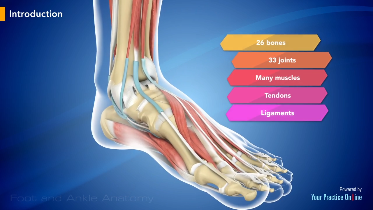
Foot And Ankle Anatomy Video Foot Ankle from www.ypo.education A muscle on the front part of the upper arm. The hip itself is a ball and socket joint, much like the shoulder.the structures necessary to create this joint are the socket, the joint capsule, muscle, ligaments, and the neck. 9 photos of the foot tendons and ligaments diagram. Black and white print showing the musculoskeletal system of a human hand, including the bones, muscles, cartilage, tendons, ligaments, and joints,. The anterior cruciate ligament prevents the femur from sliding backward on the tibia (or the tibia sliding forward on the femur). Also allows the action of raising up onto toes. The fleshy, thick part of the muscle is called its belly. If you tear the biceps tendon at the shoulder, you may lose some strength in your arm and have pain when you forcefully turn your arm from palm down to palm up.
The bones of the hip include the femur, the ilium, the ischium, and the pubis.
If you would like to learn all the parts of the foot structure, you have come to the right place. Tendons are remarkably strong, having one of the highest tensile strengths found among soft tissues. Foot anatomy diagram, foot joint diagram, foot sprain diagram, foot tendons and ligaments pain, leg tendon diagram. See anatomy pictures of the 27 bones in the hand and wrist how they are connected with tendons and muscles and the nerves that run through the. Tendon, tissue that attaches a muscle to other body parts, usually bones.tendons are the connective tissues that transmit the mechanical force of muscle contraction to the bones; The extensor tendon compartments of the wrist are six tunnels which transmit the long extensor tendons from the forearm into the hand. Contusions (bruises) tendonitis (inflammation of a tendon) sciatica (pain from the sciatic nerve) last medically reviewed on june 18, 2015. Tendon diagrams and design force vectors. Maybe you would like to learn more about one of these? When the muscles tighten (contract) arguably, the most important tendon is the achilles tendon, which allows the calf muscles to move. There are a whole range of structures e.g. Attaches the calf muscles to the calcaneus, most important muscles for running, jumping, walking etc. This chart is perfect for educating medical students or for patient…
Browse 318 hand anatomy tendons stock photos and images available, or start a new search to explore more stock photos and images. The tendon travels along the inside of the forearm on the side of the small finger and crosses the wrist. Contusions (bruises) tendonitis (inflammation of a tendon) sciatica (pain from the sciatic nerve) last medically reviewed on june 18, 2015. The hand incorporates countless muscles, bones, tendons and ligaments into simple motion and this chart covers them all. Ligaments join the knee bones and provide stability to the knee:
Ankle Anatomy Be In Motion Physiotherapy from www.beinmotion.ca Contusions (bruises) tendonitis (inflammation of a tendon) sciatica (pain from the sciatic nerve) last medically reviewed on june 18, 2015. A thick, triangular shoulder muscle. Your biceps tendons attach the biceps muscle to bones in the shoulder and in the elbow. Ligaments join the knee bones and provide stability to the knee: Foot anatomy diagram, foot joint diagram, foot sprain diagram, foot tendons and ligaments pain, leg tendon diagram. Tendons are thick bands of tissue that connect muscles to bones. If you would like to learn all the parts of the foot structure, you have come to the right place. Attaches the calf muscles to the calcaneus, most important muscles for running, jumping, walking etc.
Its muscle belly is in the forearm.
Ligaments join the knee bones and provide stability to the knee: On the other hand, the insertion is where a tendon attaches that muscle to the *more* movable bone. Browse 318 hand anatomy tendons stock photos and images available, or start a new search to explore more stock photos and images. This diagram depicts human anatomy tendons and ligaments.human anatomy diagrams show internal organs, cells, systems, conditions, symptoms and sickness information and/or tips for healthy living. Hand a hand is a prehensile multi fingered appendage located at the end of the forearm or forelimb of primates such as humans chimpanzees monkeys and lemurs human anatomy for the artist the dorsal hand the dorsal the easiest tendons to identify in the dorsal hand are those of the extensor digitorum muscle its name means extensor of the digits which is Foot anatomy diagram, foot joint diagram, foot sprain diagram, foot tendons and ligaments pain, leg tendon diagram, peroneal tendonitis, foot, foot anatomy diagram, foot joint diagram, foot sprain diagram, foot tendons and ligaments pain, leg tendon diagram, peroneal tendonitis. The tendon travels along the inside of the forearm on the side of the small finger and crosses the wrist. The achilles tendon connects the heel to the calf muscle and is essential for running jumping and standing on the toes. Muscle strains (pulls or tears) muscle cramps; Tendon, tissue that attaches a muscle to other body parts, usually bones.tendons are the connective tissues that transmit the mechanical force of muscle contraction to the bones; See anatomy pictures of the 27 bones in the hand and wrist how they are connected with tendons and muscles and the nerves that run through the. The hand incorporates countless muscles, bones, tendons and ligaments into simple motion and this chart covers them all. The anterior cruciate ligament prevents the femur from sliding backward on the tibia (or the tibia sliding forward on the femur).